Di Team (Department of Implant Dentistry)
Our long-term goal is to advance the science of dental implantology by developing innovative biomaterials, regenerative strategies, and precision treatment technologies to improve peri-implant tissue health and long-live implant success.
Our laboratory focuses on exploring novel materials and biological approaches to enhance osseointegration and promote long-term stability of dental implants. We utilize both in vitro systems and in vivo pre-clinical animal models to systematically evaluate the biological performance and clinical translational potential of newly developed therapies.

The first major research thrust centers on designing biofunctionalized implant surfaces and smart coatings by integrating bioinspired materials, such as mussel-inspired polydopamine, antibacterial agents, and osteoinductive factors. Our aim is to optimize the biological response at the bone-implant interface and enhance early and long-term osseointegration, especially under compromised conditions.
The second major research thrust focuses on advancing digital technologies in implant dentistry. We are developing and optimizing fully digital workflows for implant planning, guided surgery, and prosthetic rehabilitation. Our research involves the integration of cone-beam computed tomography (CBCT), intraoral scanning, computer-aided design and manufacturing (CAD/CAM), and 3D printing to achieve highly accurate, personalized, and efficient implant treatments. Additionally, we are exploring the application of artificial intelligence (AI) and machine learning algorithms to enhance diagnostic accuracy, predict implant outcomes, and support clinical decision-making in complex cases.
The third major research thrust addresses the mechanical and biological challenges in full-arch implant-supported prostheses. We are investigating novel digital workflows, advanced biomaterials, and biomechanical designs to optimize the precision, durability, and biological compatibility of full-arch restorations.
DIRECTOR

Ping Di, DMD
Professor
Principal Investigator
diping@bjmu.edu.cn
Professor Di graduated and remained at Peking University School and Hospital of Stomatology in 1991. She began her specialization in implant dentistry in 1995 and obtained her master’s degree in Stomatology from Peking University in 2001. In 2002, she received systematic and standardized training in implant dentistry in Germany. From 2009 to 2010, she served as a visiting scholar at the University of Tübingen, Germany, under the mentorship of the internationally renowned Professor Weber, where she completed advanced clinical training and research, earning her clinical doctorate degree.
Professor Di has long been dedicated to both clinical practice and fundamental research, with a focus on integrating modern implantology theories, cutting-edge diagnostic and therapeutic technologies, and a patient-centered medical humanities approach to continuously refine clinical implant practices. She has published many professional articles in implantology, led multiple national and provincial-level research projects, and contributed to 12 textbooks and professional monographs. As a principal investigator, she has been awarded the Second Prize of the Chinese Medical Science and Technology Award and the First Prize of the Chinese Stomatological Association Science and Technology Award. Individually, she has received multiple awards for Outstanding Clinical Service at the Peking University School and Hospital of Stomatology.
Professor Di currently holds several important academic positions, including Deputy Director of the National Clinical Research Center for Oral Diseases Implantology Alliance, Deputy Chair of the Implant Dentistry Committee of the Beijing Stomatological Association, Standing Committee Member of the Aesthetic Dentistry Committee of the Beijing Stomatological Association, and Committee Member of the Implant Dentistry Committee of the Chinese Stomatological Association. She also serves as the Associate Editor of the Chinese edition of Clinical Implant Dentistry and Related Research (CIDR) and the Chinese edition of the Journal of Oral and Maxillofacial Implants (JOMI).
RESEARCH AREAS
Infectious Bone Regeneration
Di Team is committed to developing innovative regenerative strategies for infectious alveolar bone defects. We have engineered a multifunctional scaffold by incorporating silver nanowires into an intrafibrillar mineralized collagen matrix (IMC/AgNWs), achieving both strong antibacterial effects and enhanced osteogenic performance. IMC/AgNWs demonstrate broad-spectrum antimicrobial activity against key periodontal pathogens such as Porphyromonas gingivalis and Streptococcus mutans, while promoting the osteogenic differentiation of periodontal ligament stem cells under both inflammatory and non-inflammatory conditions. In preclinical models of infected periodontal and peri-implant defects in rats and beagle dogs, the scaffolds significantly enhanced vertical bone regeneration and osseointegration through activation of the BMP/Smad signaling pathway. This work highlights the promising translational potential of IMC/AgNWs and provides a new therapeutic approach for managing infectious vertical bone defects in clinical implantology.
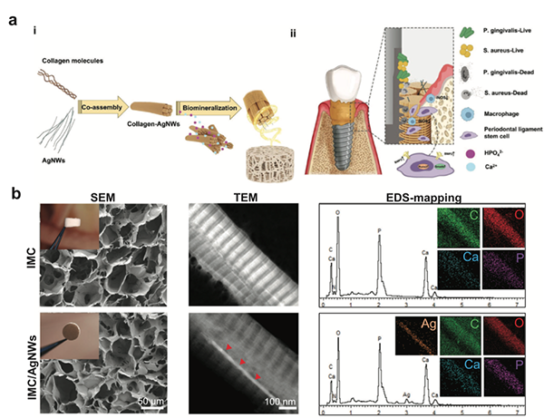
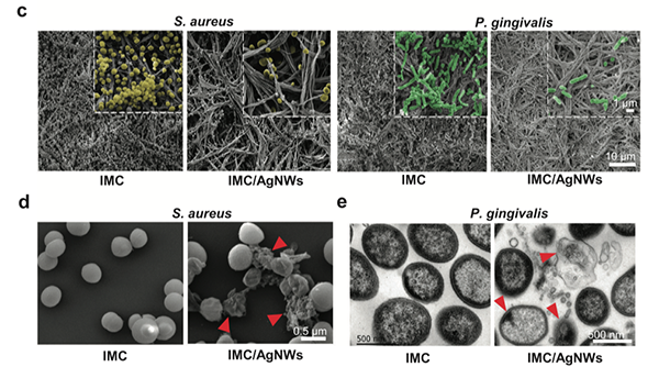
Physicochemical characterization and biocompatibility of IMC/AgNWs. a) (i) Schematic diagram of the assembly of IMC/AgNWs and (ii) the regenerative process of peri-implantitis by IMC/AgNWs in beagle dogs. b) SEM, TEM, and EDS element mapping of IMC/AgNWs. Inset in SEM images: general views of scaffolds. The silver element could be clearly identified in EDS curves and mapping. c) Representative SEM images of the micro-morphology of S. aureus (yellow, 24 h) and P. gingivalis (green, 72 h) cultured on IMC and IMC/AgNWs. d) Representative SEM images of supernatant of S. aureus cultured with IMC and IMC/AgNWs (right) for 24 h. The membranes in the IMC/AgNWs group were broken and dissolved (red arrows). Scale bar = 1 μm
Digital Occlusal Contact Analysis
Di Team focuses on advancing digital technologies in occlusal diagnostics. We have established an innovative method to compare occlusal contact regions (OCRs) captured by intraoral scanning systems and conventional impression procedures. In a study involving participants with stable centric occlusion, intraoral scanning demonstrated significantly higher consistency (0.73) and improved occlusal tightness compared with traditional impressions. The intraoral scanner provided more accurate occlusal records with reduced mean occlusal clearance and closer intercuspation. Our findings highlight that digital intraoral scanning offers a more reliable and objective approach for recording posterior quadrant occlusion. Furthermore, by quantifying occlusal clearance, we propose occlusal tightness as a critical and indispensable parameter in evaluating virtual occlusal records, offering a new direction for improving precision in clinical prosthodontics.
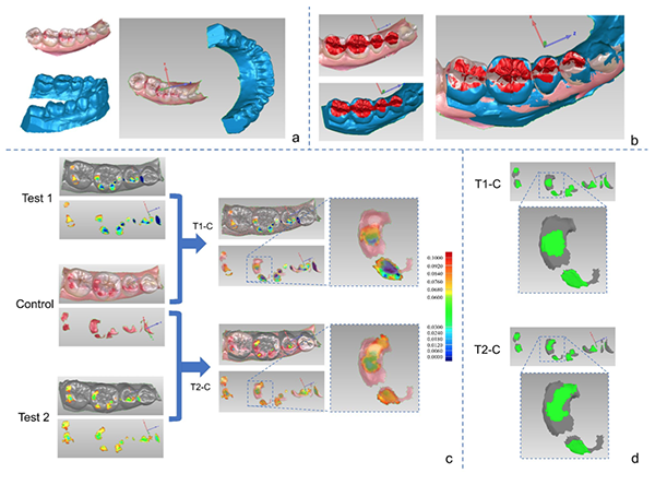
Three-dimensional superimposition of occlusal contact regions (OCRs) between the control and test groups. a The digital models were imported into the reverse engineering software. The occlusal records obtained by articulating paper were visualized on the digital models obtained from intraoral scans. b The digital models of control group and test groups were superimposed using “best-fit alignment” based on the tooth surfaces. c The distances from 0 to 100 μm between the upper and lower models were calculated and displayed on the spectrum. The three sets of images on the left side, from top to bottom, represented the OCRs of the T1 group, control group, and T2 group, respectively. The two sets of images on the right side, from top to bottom, depicted the three-dimensional comparison of OCRs between the T1 group and control group, as well as between the T2 group and control group. d The overlapping OCRs were displayed in green color. The two sets of images, from top to bottom, respectively depicted the overlapping OCRs between the T1 group and control group, and the overlapping OCRs between the T2 group and control group
Materials Performance in CAD-CAM
Di Team has made significant contributions in the field of digital restorative materials, particularly in the analysis of color changes in CAD-CAM monochromatic materials. In an in vitro study, we systematically evaluated the color variation of six different CAD-CAM monochromatic materials with varying thicknesses and surface roughness. Using the CIELab color system, we investigated the impact of material type, thickness, and surface treatments on color parameters (L*, a*, b*) and measured them with a spectrophotometer and profilometer. The results showed that material type, thickness, and surface roughness are key factors influencing color change. As thickness increased, there were significant changes in the L*, a*, and b* values, while roughened surfaces exhibited more noticeable color differences, often exceeding clinically acceptable thresholds. These findings provide important insights into the optimization of CAD-CAM monochromatic materials and offer scientific guidance on how to avoid undesirable color variations in clinical applications.
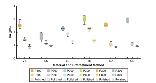
Mean and standard deviation of surface roughness after different pretreatment methods. CD, Celtra Duo; LS, IPS e.max; LU, LAVA Ultimate; TE, Telio CAD; VE, VITA Enamic; VS, VITA Suprinity
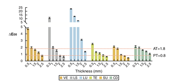
Mean and standard deviation of ΔE00 between polished specimens and those roughened with P800-grit. CD, Celtra Duo; LS, IPS e.max; LU, LAVA Ultimate; TE, Telio CAD; VE, VITA Enamic; VS, VITA Suprinity
RECENT PUBLICATIONS
A biomimetic multifunctional scaffold for infectious vertical bone augmentation
Advanced Science, 2024, 11(26): 2310292.
The facial–coronal ridge crest alterations after single immediate implant placement and provisionalization with thin buccal plate phenotype in anterior maxilla: A radiographic case‐series study
Clinical Implant Dentistry and Related Research, 2024, 26(2): 317-326.
Impact of labially inclined implant axes on immediate implant placement and provisionalization in anterior maxilla: A prospective cohort study
Clinical Implant Dentistry and Related Research, 2024, 26(6): 1200-1208.
Quantitative analysis of the color in six CAD-CAM dental materials of varied thickness and surface roughness: An in vitro study
The Journal of Prosthetic Dentistry, 2024, 131(2): 292. e1-292. e9.
Digital registration versus cone-beam computed tomography for evaluating implant position: a prospective cohort study
BMC Oral Health, 2024, 24(1): 304.
Crown adjustment and chairside efficiency of single‐unit restorations fabricated from immediate and staged impressions using a digital workflow for posterior implants
Journal of Prosthodontics, 2024, 33(7): 637-644.
Interface misfit of conventionally milled and novel hybrid full-arch implant-supported titanium frameworks
BMC Oral Health, 2024, 24(1): 1205.
An innovative evaluation method for clinical comparative analysis of occlusal contact regions obtained via intraoral scanning and conventional impression procedures: a clinical trial
Clinical Oral Investigations, 2024, 28(10): 543.
The impact of mandibular partial edentulous distal extension on virtual occlusal record accuracy when using two different intraoral scanners: An in vitro analysis
Journal of Dentistry, 2024, 150: 105303.
Lateral sinus floor elevation in patients with sinus floor defects: A retrospective study with a 1‐to 9‐year follow‐up
Clinical Oral Implants Research, 2023, 34(10): 1141-1150.
Comparison of 4‐or 6‐implant supported immediate full‐arch fixed prostheses: A retrospective cohort study of 217 patients followed up for 3–13 years
Clinical Implant Dentistry and Related Research, 2023, 25(2): 381-397.
A Novel Method to Achieve Preferable Bone-to-Implant Contact Area of Zygomatic Implants in Rehabilitation of Severely Atrophied Maxilla
International Journal of Oral & Maxillofacial Implants, 2023, 38(1).
Plaque accumulation on the fitting surface of full‐arch implant‐supported fixed prostheses with contact or noncontact pontics: A split mouth randomized controlled trial
Journal of Esthetic and Restorative Dentistry, 2023, 35(7): 1077-1084.
Comparison of 4‐or 6‐implant supported immediate full‐arch fixed prostheses: A retrospective cohort study of 217 patients followed up for 3–13 years
Clinical Implant Dentistry and Related Research, 2023, 25(2): 381-397.
Effects of thickness and polishing treatment on the translucency and opalescence of six dental CAD-CAM monolithic restorative materials: an in vitro study
BMC Oral Health, 2023, 23(1): 579.
Clinical evaluation and quantitative occlusal change analysis of posterior implant‐supported all‐ceramic crowns: A 3‐year randomized controlled clinical trial
Clinical Oral Implants Research, 2023, 34(11): 1188-1197.
Quantitative analysis of color accuracy and bias in 4 dental CAD-CAM monolithic restorative materials with different thicknesses: An in vitro study
The Journal of Prosthetic Dentistry, 2022, 128(1): 92. e1-92. e7.
Accuracy and feasibility of 3D-printed custom open trays for impressions of multiple implants: A self-controlled clinical trial
The Journal of Prosthetic Dentistry, 2022, 128(3): 396-403.
Dental implant nano-engineering: advances, limitations and future directions
Nanomaterials, 2021, 11(10): 2489.
MiR-181d-5p regulates implant surface roughness-induced osteogenic differentiation of bone marrow stem cells
Materials Science and Engineering: C, 2021, 121: 111801.
TEAM
Shuai Li, DMD
Researcher
lisengbj@qq.com

Shuxin Ren, DMD
Researcher
woodbubu@163.com
Jiehua Tian, DMD
Researcher
Tianjiehua8866@163.com

Xiaosong Yi, DMD
Researcher
kqyixs@163.com
Donghao Wei, DMD
Researcher
Weidh_pku2011@163.com

Yifan Zhang, DMD
Researcher
Zyfbjmu2012@163.com

Zhengda Wu, DMD
Postdoctoral Researcher
chairgotcold@163.com
CONTACT
Ping Di
diping@bjmu.edu.cn
+86-10-82195344
Department of Implant Dentistry,
Peking University School and Hospital of Stomatology,
No.22 Zhongguancun South Avenue,
Haidian District, Beijing 100081, PR China
last text: Lin Team (Department of Implant Dentistry)
next text: Yuming Zhao Lab


